Development of Radiofrequency Microcoils Towards Oxygen Guided Radiation Treatment
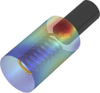
Tumor hypoxia (low oxygen) is a major adverse factor in cancer that leads to poor outcomes regardless of treatment modality. Hypoxia was demonstrated to significantly correlate with increased radiation resistance, treatment failure, and metastasis. Pre-clinical experiments where the dose of radiation therapy was adjusted based on the local tissue oxygen levels obtained via electron paramagnetic resonance (EPR) imaging demonstrated the critical role of hypoxia in treatment outcomes. However, there is no clinical method or device capable of measuring tissue oxygen levels in real-time under clinical conditions. We present the development of four EPR radio frequency micro coils (RFMC) designed for clinical use to enable personalization of radiation treatments based on the oxygen levels at different locations inside tumor tissues. The purpose of the RFMCs is to provide an oscillating magnetic field (B1) to the oxygen sensitive spin probe composed of lithium phthalocyanine (LiPc) and medical grade silicone to excite the free electrons in the probe. All RFMC designs were attached to a miniature (1.2mm outer diameter) coaxial transmission line (UT-047) with 0.40 dB/foot attenuation. The RFMC designs we investigated are: 1) a multi-turn micro coil, 2) a skewed helical micro coil, 3) a coplanar micro strip, and 3) a single turn closed circuit loop. Simulations of ?1⊥ field distributions arising from the RFMC designs were conducted using the radio frequency (RF) module in the commercial finite element analysis software COMSOL Multiphysics. For each RFMC design, the oscillating magnetic field perpendicular to the main magnetic field (B1) was plotted on a parametrized cylindrical surface with a small OD (1.98mm) representing the LiPc probe (Figure 1). Surface integration of 〖B1〗_⊥ was performed along the LiPc cylinder to allow comparison across all RFMC designs. 2D polar plots of the radiation pattern in the x-y plane were created to compare the field gain around the HDR needle circumference (Figure 2). To calculate the active volume of each RFMC, we plotted the normalized ?1⊥^2 values along the x, y, and z coordinates and the active volume was defined as the distance between boundaries where B1 dropped significantly (by 80%). The simulation results showed that the skewed helix produced the strongest oscillating ?1⊥ field and its active volume had the greatest coverage of the LiPc probe location. The RFMC geometries were defined using parametric equations to allow parameter sweep studies varying the micro coil dimensions. We will expand our parametric sweep simulations allowing orientation, size, and materials of RFMC components to vary within the seeking to maximize the strength of the ?1⊥ field, the overlap of the active volumes with the oxygen sensitive LiPc probe layer on the outer HDR needle walls, and the alignment of each RFMC’s directionality in the H-plane with the spatial distribution of distinct LiPc regions of interest. Future work will include optimization of the power-to-field conversion efficiency.

